A young woman presented with a several month history of a fullness in her abdomen associated with pain. Upon examination, one could palpate a ball-like mass in her pelvis. It wasn’t stuck in one place, however. It moved slightly when pressed. This sign usually reveals that the mass is either attached to the ovaries or to the intestines, the former being much more common.
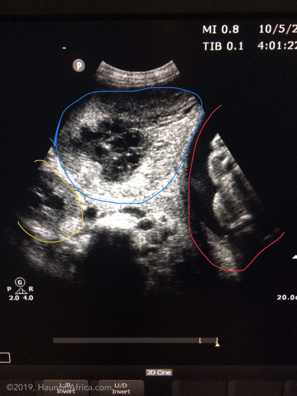
On ultrasound, the mass had multiple compartments that were a mix of patterns. It wasn’t uniform. The patient appeared otherwise healthy. We discussed her options for treatment and that only through surgery could we fully diagnose the assumed mass on her ovary, and that there was a chance it could be a cancer.
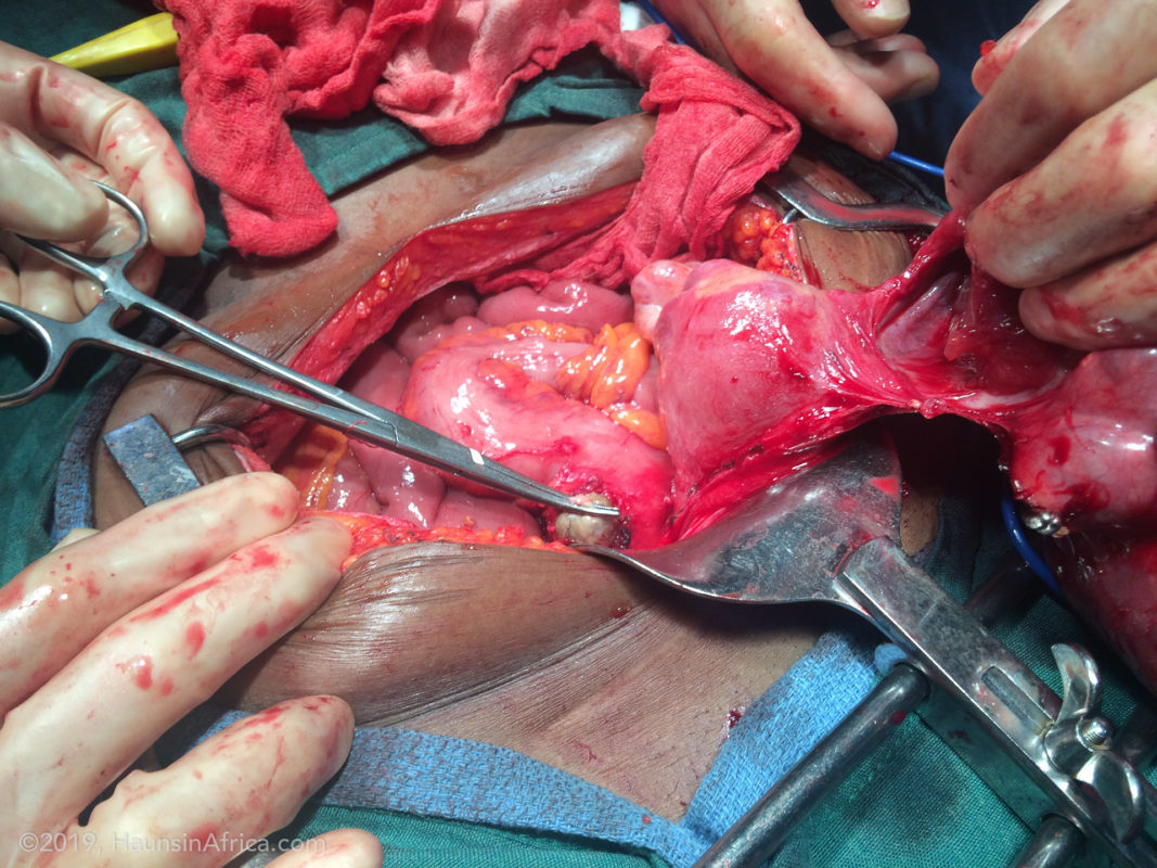
In the operating room, we found a large cyst that had engulfed her right ovary. The left appeared to be normal. As we continued to free this tumor from the surrounding structures, we had difficulty freeing it from the rectum. We finally realized that a small portion of the mass had actually started to perforate the rectum and it was growing inside the rectal wall!!! We were able to remove both the large and small tumors. The small defect in the rectal wall was repaired in two layers.


The patient did very well postoperatively.
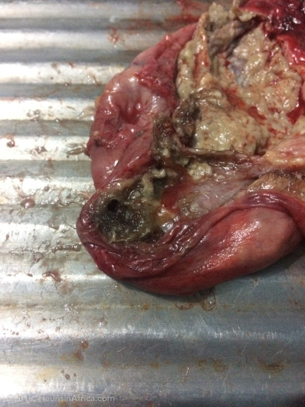
The specimen was opened up on the back-table. It revealed hairballs, bony hard nodules and large pockets of secretions (especially sebaceous fluid which is thick, sticky and foul smelling). The smaller tumor was just a cyst of hardened sebaceous fluid. The final pathology was a mature cystic teratoma or dermoid cyst of the right ovary. It is a benign tumor, meaning that it is not cancer. Removing the tumor was the only treatment needed.